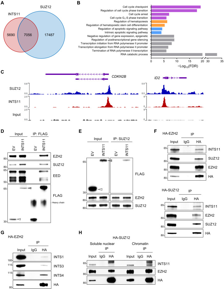
Fig. 5. INTS11 forms a complex with PRC2.
(A) Venn diagram showing the overlap among binding sites (peaks) of INTS11 (GSE106359) and SUZ12 (GSE59090) in hematopoietic cells. (B) Gene ontology analysis of the genes that were co-occupied by INTS11 and SUZ12. (C) Representative ChIP-seq tracks show that INTS11 and SUZ12 co-occupy PRC2 target gene promoters in hematopoietic cells. (D) Nuclear extracts of 293T cells transfected with FLAG-tagged INTS11 or empty vector control (EV) were immunoprecipitated (IP) with anti-FLAG and probed for PRC2 proteins. Arrowhead indicates the FLAG-fusion protein. (E) Reciprocal SUZ12 IP from nuclear extracts of 293T cells transfected with FLAG-tagged INTS11 or EV, and representative immunoblot analysis. (F) Nuclear extracts of 293T cells transfected with HA-tagged EZH2 (top) or SUZ12 (bottom) were IP with anti-HA or mouse IgG, and Western blotting was performed. (G) Western blot analysis of the samples above (F) using antibodies against INT subunits INTS1, INTS3, and INTS4. (H) Soluble nuclear and chromatin-bound fractions from 293T cells transfected with HA-tagged SUZ12 were subjected to IP with anti-HA antibody followed by Western blot analysis with indicated antibodies.

