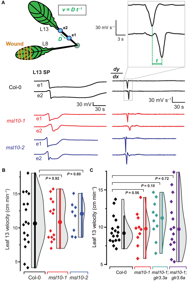
Fig. 4. L13 SWP velocities are unaffected.
(A) Schematic showing of wounded L8 and placement of two electrodes (e1 and e2, blue dots) on the petiole of distal L13. Example SWP recordings are shown below the schematic, and the derivative (dy/dx) is shown to the right. Velocity (v) was calculated as the distance (D) between two electrodes (e1 and e2) divided by the time (t) between derivative minima. (B) Quantification of velocity of SWP in msl10 mutants versus Col-0 (median and SEM: Col-0: 10.7 ± 1.0 cm min−1, msl10-1: 10.9 ± 0.8 cm min−1, msl10-2: 12.9 ± 0.8 cm min−1). (C) Quantification of SWP velocity in L13 of msl10-1 single mutant or msl10-1;glr3 double mutants in comparison to Col-0. Violin plots and statistics as in Fig. 1 (n = 8 to 17). All P values calculated by Mann-Whitney U test.

