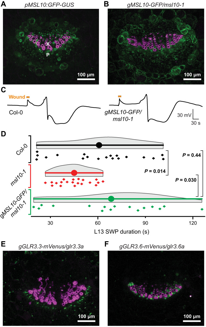
Fig. 5. Expression of GFP-tagged transcriptional and translational MSL10 in leaf vascular bundles.
(A) Composite image of a transverse petiole section showing fluorescence from a GFP-GUS transcriptional reporter fused to MSL10 promoter (pMSL10) in green and lignin autofluorescence indicating xylem vessels in magenta. Image is a maximum intensity z-stack projection of confocal data. X, xylem; P, phloem. (B) Composite image of a transverse petiole section showing fluorescence from a MSL10 genomic fragment (gMSL10) tagged with GFP in green and lignin autofluorescence indicating xylem vessels in magenta. GFP-tagged construct is expressed in the msl10-1 mutant background. (C) Representative SWP traces from an experiment comparing Col-0 plants to msl10-1 mutants transformed with translational gMSL10-GFP reporter construct. (D) Statistical analysis of an independent experiment comparing Col-0, msl10-1, and gMSL10-GFP/msl10-1 SWP durations in L13. Displayed P values calculated by Mann-Whitney U test (n = 16 to 19). Statistical analysis of a third independent experiment is shown in fig. S11. (E and F) Composite images of transverse petiole sections of gGLR3.3-mVenus and gGLR3.6-mVenus translational reporters expressed in cognate mutant backgrounds showing mVenus fluorescence in green and lignin autofluorescence in magenta. Refer to fig. S11 for individual fluorescence channels and analysis of an independent complementation line.

