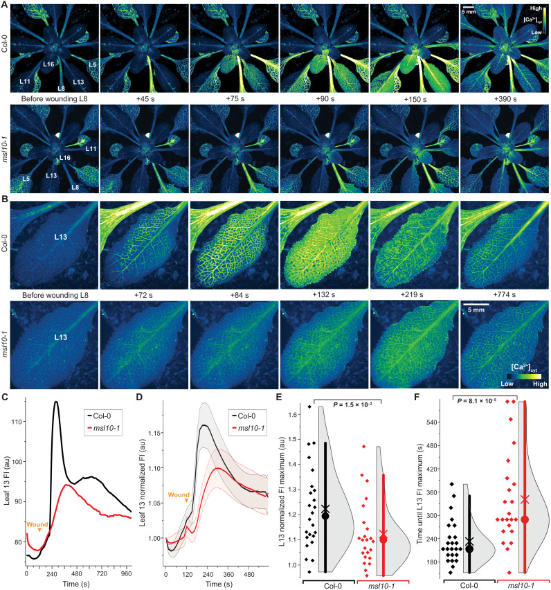
Fig. 7. Quantitative imaging of wound-induced cytosolic calcium ([Ca2+]cyt) dynamics in adult plants using MatryoshCaMP6s.
(A) Whole-plant imaging reveals preferential spread of cytosolic calcium elevations to neighboring parastichous leaves upon wounding L8. Calcium elevations in leaves parastichous to L8 are attenuated in msl10-1 mutant background. (B) Close-ups of L13 calcium imaging in Col-0 and msl10-1 plants. (C) Time course quantification of data shown in (B), showing raw fluorescence intensity (FI) in arbitrary units (au). (D) Averaged FI, normalized to initial time point (FIt/FI0), observed in L13 of Col-0 and msl10-1 plants (n = 23 to 24; error bands, SEM). (E and F) Quantification and statistical analyses of L13 normalized FI maxima and time until maxima after wounding. Bar, 1.5× interquartile range; circle, median; ×, mean. Displayed P values were calculated by Mann-Whitney U test (n = 23 to 24). For more detailed spatiotemporal information, refer to fig. S13 and movies S1 to S4.

