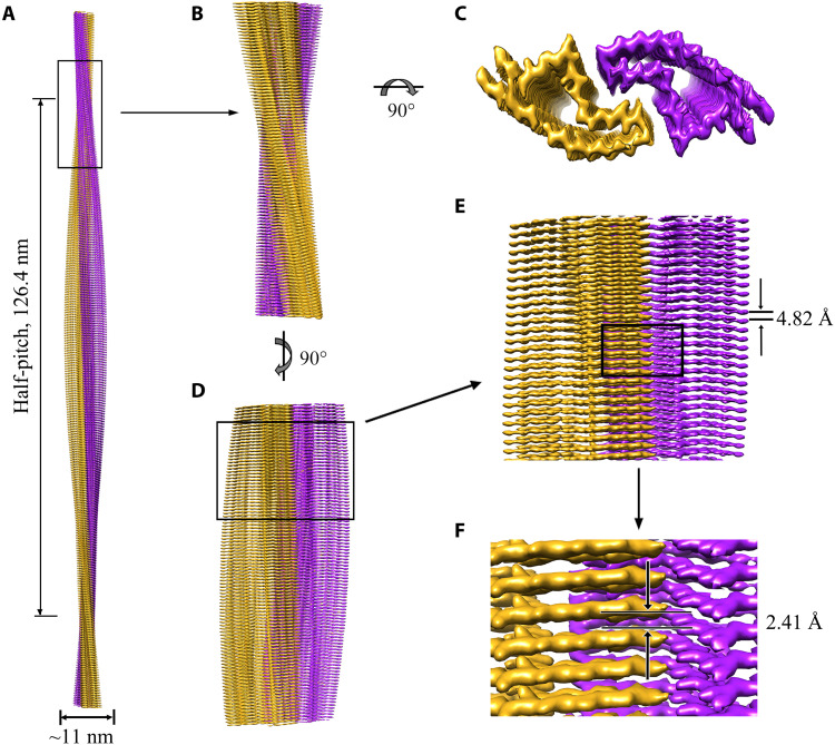
Fig. 2. Cryo-EM structure of E196K fibrils.
(A) 3D map showing two protofibrils intertwined into a left-handed helix, with a fibril core width of ~11 nm and a half-helical pitch of 126.4 nm. The two intertwined protofibrils are colored purple and gold, respectively. (B) Enlarged section showing a side view of the density map. (C) Top view of the density map. (D) Another side view of the density map after 90° rotation of (B) along the fibril axis. (E) Close-up view of the density map in (D) showing that the subunits in each protofibril stack along the fibril axis with a helical rise of 4.82 Å. (F) Close-up view of the density map in (E) showing that the subunits in two protofibrils stack along the fibril axis with a helical rise of 2.41 Å.

