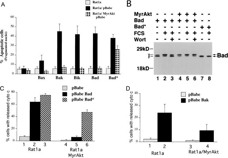
FIG. 6.
Akt inhibits apoptosis and cytochrome c release induced by overexpression of the proapoptotic Bcl-2 family members. (A) Akt inhibits apoptosis accelerated by overexpression of Bax, Bak, Bik, and Bad. Rat 1a and Rat1a/MyrAkt cells were infected with eGFP retroviruses expressing Bax, Bak, Bik, Bad, and the Bad* (see Materials and Methods). Apoptosis was scored by DAPI staining 16 h following serum deprivation, a time point at which Rat 1a cells do not exhibit significant apoptosis. Cells were seeded at a density of 50,000 cells/3-cm-diameter dish. The next day cells were placed in DMEM containing 0% FCS for 16 h. Averages (± standard errors) of at least 300 cells from three independent experiments. (B) Akt induces a mobility shift of ectopically expressed Bad in Rat1a cells. Rat1a (lanes 1 to 3, 7, and 8) and Rat1a/MyrAkt (lanes 4 to 6) cells were infected with retrovirus expressing Bad or Bad*. Forty-eight hours following infection, cells were deprived of serum for 2 h. Cells were either mock treated (lanes 1, 2, 4, 5, 7, and 8) or treated with 200 nM wortmannin (Wort; lanes 3 and 6) for 30 min. Cells were then either mock treated (lanes 1, 4, and 7) or stimulated with 20% FCS for 30 min (lanes 2, 3, 5, 6, and 8). Cells were lysed, and extracts were subjected to SDS-PAGE (12% gel). Bad was detected with a hamster monoclonal anti-Bad antibody (PharMingen clone 13361S), and the different phosphorylation states are indicated with arrows. Positions of molecular weight standards are indicated on the left. (C) Akt prevents cytochrome c release accelerated by overexpression of Bad. Rat1a (columns 1 to 3) and Rat1a/MyrAkt (columns 4 to 6) cells demonstrating diffuse cytochrome c immunostaining were quantitated. Cells were treated as described for Fig. 2. Two hours (a time point at which there is no significant cytochrome c release in Rat 1a cells) after UV irradiation cells were fixed and immunostained for cytochrome c localization. Averages (± standard errors) of at least 100 cells from three independent experiments are shown. (D) Akt prevents cytochrome c release accelerated by overexpression of Bak. Cells were treated and scored for cytochrome c release as described for panel C.

