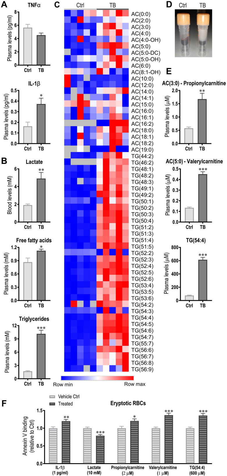
Fig. 4. Tumor-induced metabolic remodeling and immune activation promotes eryptosis.
(A and B) Levels of proinflammatory cytokines TNFα and IL-1β (A) and blood lactate, free fatty acids, and triglycerides (B) were analyzed in plasma of Ctrl and TB mice 3.5 weeks after tumor inoculation. (C) Heatmap of acylcarnitines (ACs) and triglycerides (TGs) from the metabolomics analysis. (D) Representative picture of plasma of Ctrl and TB mice demonstrating the massive elevation of lipids. (E and F) The effects of candidates identified in metabolomics analysis (E) and IL-1β and lactate on eryptosis were tested by incubating healthy RBCs with the observed levels of IL-1β or the different metabolites. After 24 hours, eryptosis was measured by assessing phosphatidylserine externalization via annexin V binding using flow cytometry (F). Groups (n = 5 to 7 per group) were compared by two-tailed unpaired t tests with Welch’s correction and data are represented as means + SEM. Data of in vitro eryptosis experiments consist of three independent experiments with three to four biological replicates. Asterisks indicate differences between Ctrl and TB/treated group. *P < 0.05, **P < 0.01, and ***P < 0.001. Photo credit: Regula Furrer, University of Basel.

