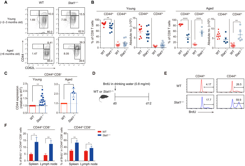
Fig. 1. Stat1−/− mice exhibit an enhanced number of CD44hi MP CD8+ T cells.
(A) Flow cytometry for CD44 and CD62L expression in splenic CD8+ T cells from young versus aged WT and Stat1−/− mice. (B) Percentage and number of CD44lo and CD44hi CD8+ T cells from WT and Stat1−/− mice. (C) Relative levels of CD44 expression in WT versus Stat1−/− CD44lo CD8+ T cells. (D) Experimental scheme of in vivo BrdU uptake. (E and F) BrdU incorporation of CD44lo and CD44hi CD8+ T cells from WT and Stat1−/− mice. The results are presented as means ± SEM. Data are representative of three to four independent experiments. *P < 0.05, **P < 0.01, and ****P < 0.0001.

