
Figure 1: Multi-contrast and parametric CMR imaging in patient with confirmed ICI myocarditis (1.5T).
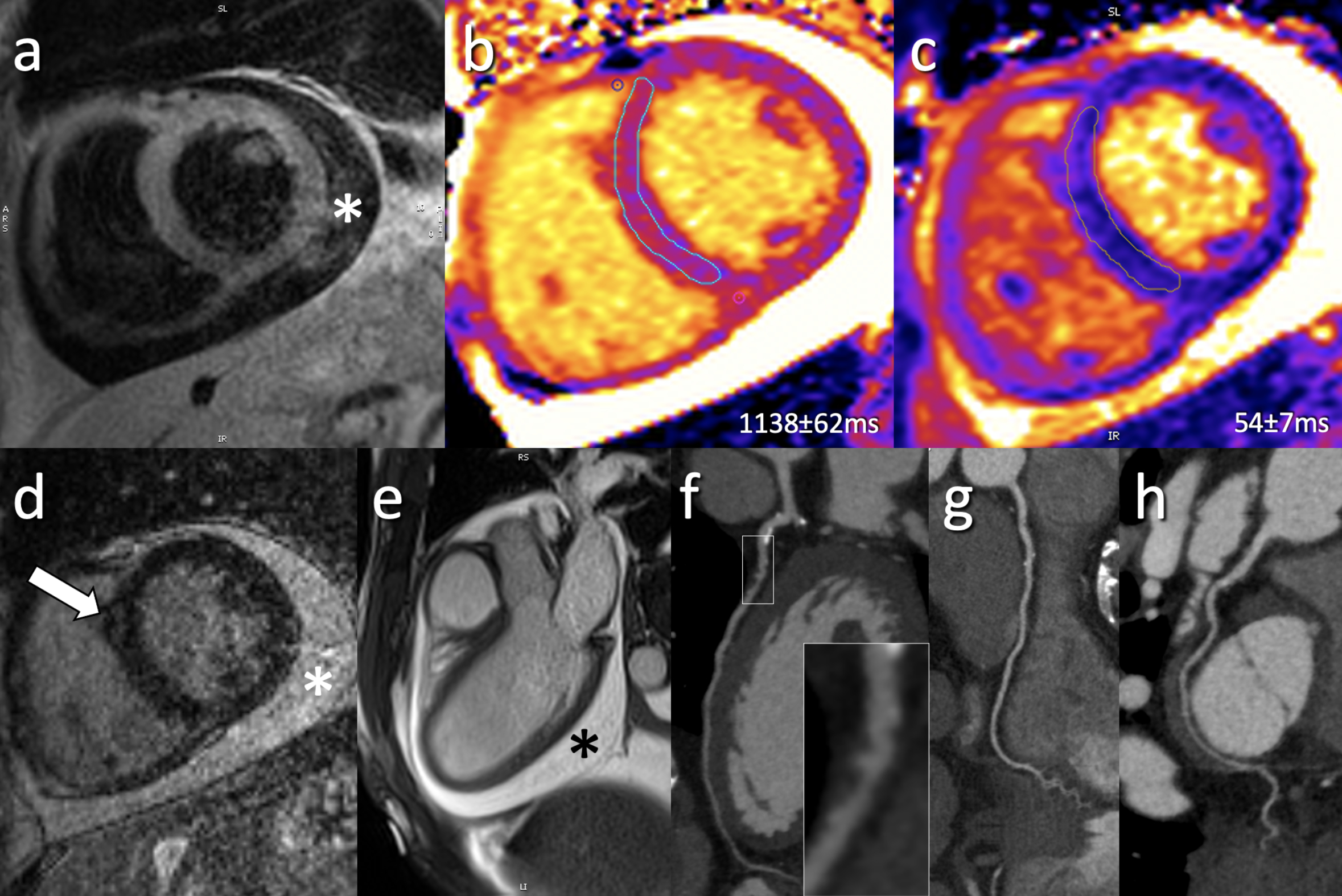
(a) Representative short axis T2-weighted SPAIR without focal signal abnormality; (b) Mid-ventricular short axis MOLLI T1-map demonstrates diffusely elevated T1 values (local normal reference: 1006±24ms) while (c) same slice T2-map demonstrate normal global T2 values (local normal reference: 52±3ms); (d) Post-contrast LGE imaging demonstrates faint mid-myocardial enhancement (arrow) in the mid-ventricular anteroseptum; (e) 3-chamber cine bSSFP image demonstrates a pericardial effusion (*); (f-h) coronary CT angiography performed for exclusion of possible coronary artery disease demonstrated calcified and non-calcified changes (predominately in LAD; f) without significant coronary artery stenosis.