
Fig. 5.
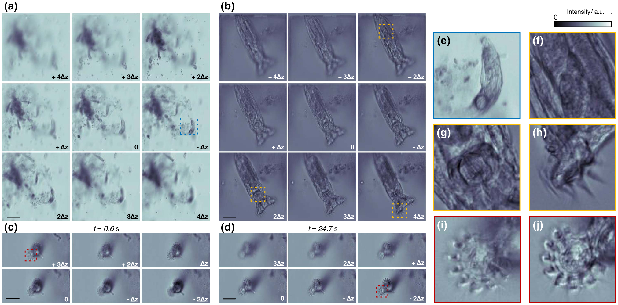
Multifocus phase-contrast imaging of living rotifers. (a) Large FOV (1.1 × 1.1 × 0.7 mm3) imaging of multiple rotifers in their natural state. Δz = 88.6 μm. Scale bar, 200 μm. (b) Multifocus image of a single rotifer at high resolution. Δz = 5.5 μm. Scale bar, 50 μm. (c), (d) High speed imaging (100 Hz) of beating cilia of a rotifer. (c) and (d) show two frames at times t = 0.6 s and t = 24.7 s, respectively. Δz = 5.5 μm. Scale bar, 50 μm. (e) Expanded view of a single rotifer within the blue square in (a). (f)–(h) Expanded view of stomach, mastax, and corona regions [yellows squares at depths +2 Δz, −2 Δz, and −4 Δz in (b)] of a rotifer at different depths. (i), (j) Expanded view of cilia from the red square regions in (c), (d), respectively. The focal position of the cilia moved from +3 Δz at t = 0.6 s to −2 Δz at t = 27.4 s. a.u., arbitrary unit.