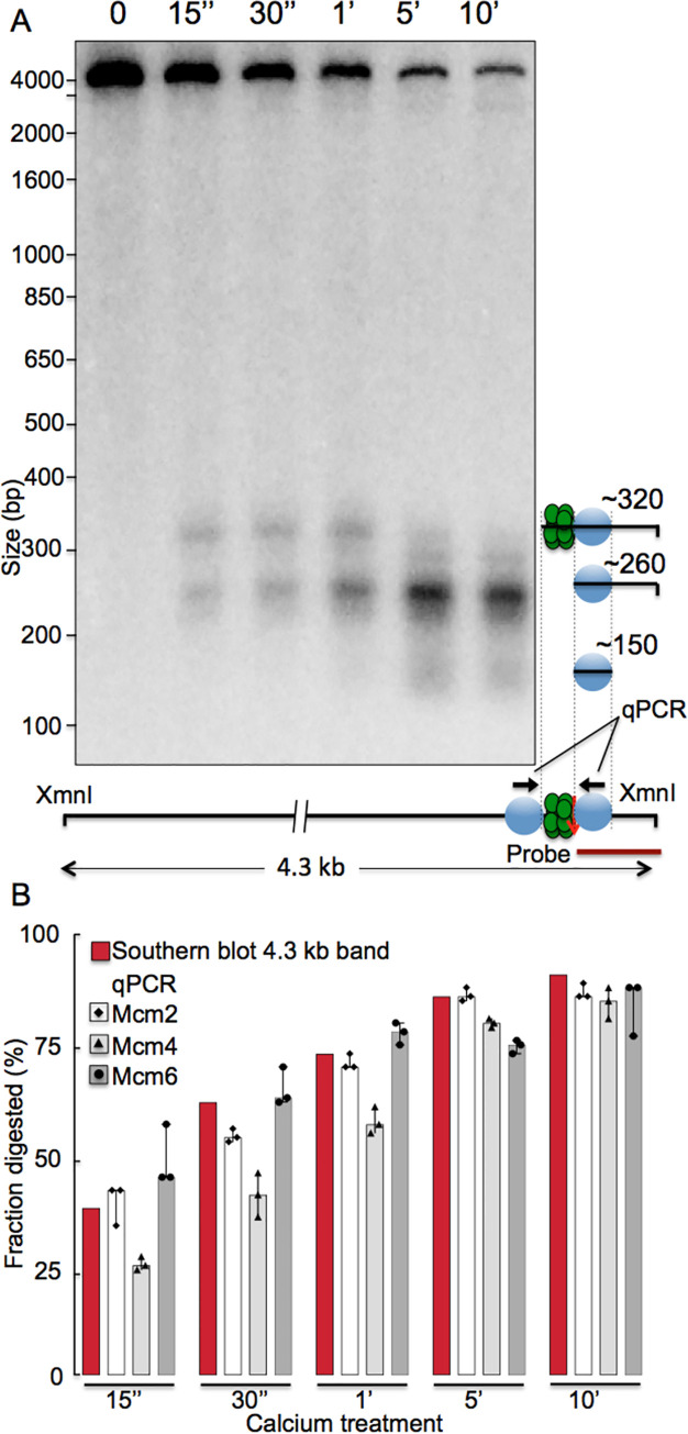
Fig 3. Kinetics of MNase cutting by Mcm-MNase fusion proteins in budding yeast, as assessed by Southern blot and qPCR.
A. S. cerevisiae cells whose endogenous copy of MCM2 was fused to MNase were permeabilized and incubated with calcium to activate the MNase for various times (listed at top). DNA was then isolated, cut with XmnI, and analyzed by Southern blot, using a radioactive probe that hybridizes immediately to the right of the ACS in ARS1103, as indicated in the lower right. Cutting by MNase can be monitored by disappearance of the 4.3 kb XmnI fragment, as it is cleaved by MNase, and by concurrent appearance of shorter bands, whose identities are indicated in the diagram on the right. Green circles depict the MCM complex and light blue circles depict the nucleosomes that flank ARS1103. B. Yeast cells carrying Mcm2, Mcm4- or Mcm6-ChEC constructs at the endogenous loci were permeabilized, the MNase was activated for various times by incubation with calcium, as described above, and cutting at ARS1103 was monitored by qPCR, using primers that flank ARS1103, as depicted by arrows on the lower right. Bars indicate disappearance of the PCR product due to cutting by MNase, with disappearance of the 4.3 kb XmnI fragment in the Southern blot shown for comparison with the results in panel A.

