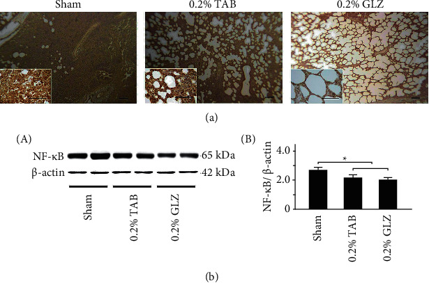
Figure 7.

GLZ treatment reduced expression of inflammation-related NF-κB in the lung tissue of weanling piglets. (a) Representative IHC stains for NF-κB expression among piglets at 10 weeks of age after sham, 0.2% TAB, and 0.2% GLZ treatments. NF-κB expression is indicated by dark brown. Scale bars are 30 μm for large images and 150 μm for small images. (b) Western blot for NF-κB expression in the lung tissue of piglets at 10 weeks of age after each treatment (n = 3 for each group). Values are mean ± standard error of the mean (∗p < 0.05, one-way analysis of variance followed by Student–Newman–Keuls multiple-comparison posttest).
