
Figure 3.
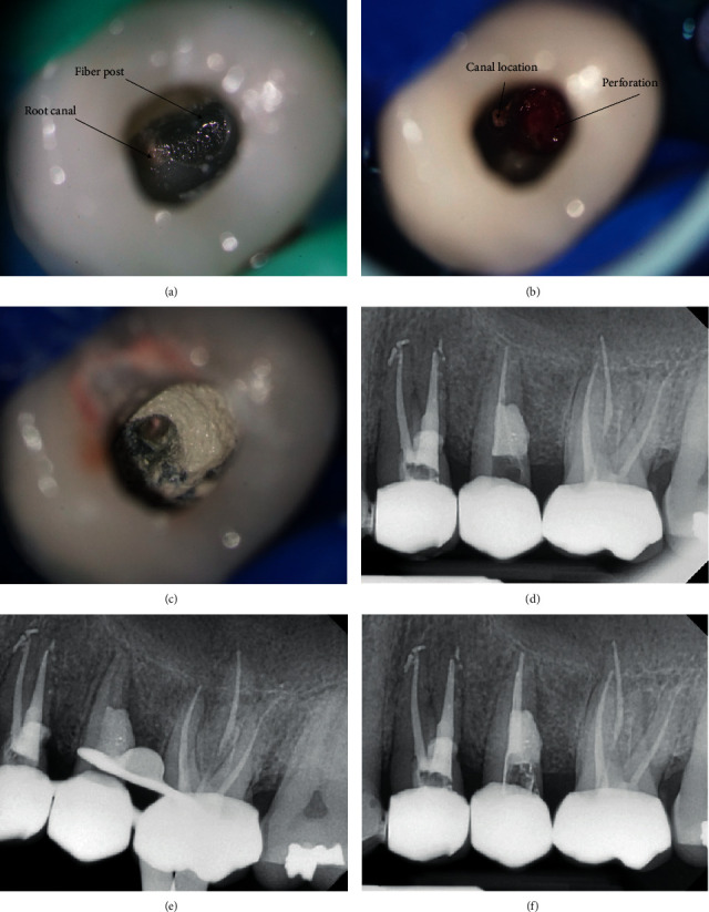
Clinical images and periapical radiographs of the retreatment of tooth #25. (a) Access cavity; (b) bleeding perforation; (c) 5MO cement application; (d) radiography of 5MO cement application; (e) radiography of cleaned and prepared canal; and (f) radiography of obturation.