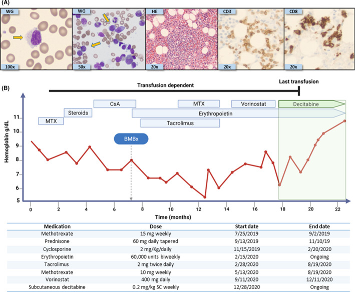
FIGURE 1.

Histopathological findings and treatment course of our index case. (A) From left to right: Wright‐Giemsa (WG) stain of peripheral blood showing a classical large granular lymphocyte (100X); WG stain of bone marrow (BM) aspiration showing classical LGL (50X); hematoxylin‐eosin stain (20X) of the BM biopsy core with infiltration of small lymphocytes characterized by CD3 (20X) and CD8 (20X) positivity by immunostaining. Flow cytometry confirmed the aberrant T‐LGL phenotype: CD2, CD3, CD5 dim, CD8, CD16, and CD57. Yellow arrows indicate large granular lymphocytes. (B) Timeline depicting the type of treatment received along with duration of administration, with dosages described underneath. MTX: Methotrexate, CsA: Cyclosporine, BMBx: Bone marrow biopsy, and SC: subcutaneously
