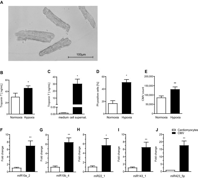
Fig. 2.
Murine cardiomyocytes were successfully isolated and cultivated from whole murine hearts visualized by light transmission microscopy (a). Increased levels of troponin-T were recorded in the supernatant of cardiomyocytes exposed to hypoxia (b). Contamination of cell culture medium with troponin-T was ruled out (c). Exposure to hypoxia led to significantly increased cardiomyocyte death compared to normoxic conditions as shown by PI staining in flow cytometry (d). More CMV were released from murine cardiomyocytes under hypoxic conditions and counted by flow cytometry (e). Murine CMV displayed a similar miRNA profile as H9c2 CMV. qPCR showed that the 5 most abundant miRNA in H9c2 CMV were also upregulated in murine CMV (black columns) compared to cardiomyocytes (white columns) (f–j)

