Fig. 2.
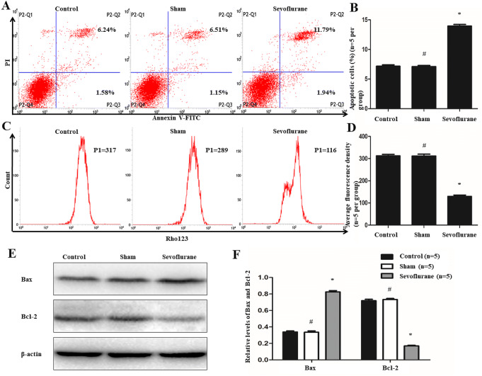
Effects of sevoflurane on the apoptosis of hippocampal neuron. a and b Flow cytometry was conducted to analysis apoptosis rates of cells, P2–Q2 (Annexin V + /PI +) and P2–Q3 (Annexin V + /PI−) represented the rates of late and early apoptosis of cells in (a), respectively. The results showed the total ratios of apoptotic cells were 7.15 ± 0.45% in Control group, 7.10 ± 0.39% in Sham group and 13.94 ± 0.55% in Sevoflurane group. Compared with the Control group, the apoptosis of cells were increased in Sevoflurane group but there were no significant changes in Sham group. c and d Flow cytometry was also used to determine the membrane potential of mitochondrial. And after treatment of sevoflurane, the fluorescence intensity of Rhodamine 123 was remarkably decreased. e and f Western blot was performed to test the levels of Bax and Bcl-2. The expression of Bax protein was elevated, while the level of Bcl-2 was attenuated after the treatment of sevoflurane. (*P < 0.01, vs Control group; #P > 0.05, vs Control group, n = 5 per group)

