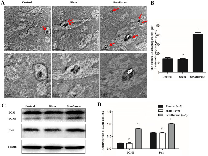
Fig. 3.
Effects of sevoflurane on autophagy and autophagic flux of APP/PS1 mice. a and b Transmission electron microscope was employed to analyze the number of autophagosomes. And the data displayed that there was a remarkable upregulation of autophagosomes at the distal axon after treatment of sevoflurane (n = 4 per group). c and d Furthermore, western blot analysis showed sevoflurane could not only enhance the levels of LC3II but also increase the expression of P62 (n = 5 per group). (*P < 0.01, vs. Control group; #P > 0.05, vs. Control group)

