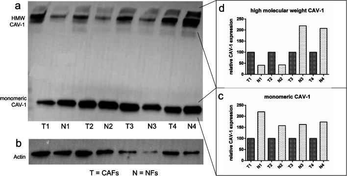
Fig. 4.
Protein expression analysis of Cav-1 in CAFs and NFs by western blotting. Here an exemplary western blot of lysates from NFs and CAFs is shown. Probes from the same patient were loaded next to each other onto the gel (T = fibroblasts for tumor tissue, N = fibroblasts from normal tissue; T1 and N1 originate from the same patient). Cav-1 could be detected as monomeric protein and as high-molecular weight fraction (a). As a reference protein, beta-actin was chosen (b). Cav-1 expression is presented in two different values, one for the monomeric form (c) and one for the high-molecular weight fraction (d). Each Cav-1 expression value was normalized with beta-actin and the paired samples from the same patient were expressed in relation to each other. This means Cav-1 expression in CAFs (= 100%) was related to Cav-1 expression in NFs of the same patient. Cav-1 expression varied between different patients in NFs and in CAFs (a). In relation to the tumor sample, the expression of monomeric Cav-1 was higher in NFs (c). In contrast, expression level of the high-molecular weight fraction of Cav-1 was heterogenous, sometimes being higher in CAFs and sometimes higher in NFs (d)

