
Figure 1.
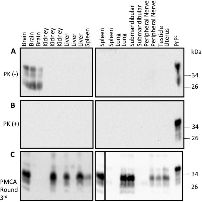
PrPC and PrPSc screening in white-tailed deer fetal tissues using western blot and PMCA. Western blot analysis of representative fetal tissue samples prior (A) and after (B) PK treatment. (C) Results from the same representative samples depicted in (A) and (B) after PMCA analysis. Numbers at the right of each panel represent molecular weight markers (in KDa). “PrPC” denotes brain extracts from Tg1536 mice not treated with PK and used as additional molecular weight markers. The solid line within panel 1C denote images
taken from different membranes.