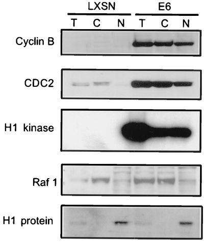
FIG. 4.
Cyclin B and CDC2 translocate into the nucleus of ADR-treated E6 cells. Asynchronously growing E6-HFFs and LXSN-HFFs were treated continuously with 100 nM ADR for 36 h. Total (T), cytoplasmic (C), and nuclear (N) proteins were harvested 36 h after exposure as described in Methods and Materials; 20 μg of total, cytoplasmic, and nuclear extracts were loaded onto an SDS–12% polyacrylamide gel and transferred. Western blot analyses for cyclin B, CDC2, the cytoplasmic control (Raf1), and the nuclear control (histone H1) were performed. The ADR-treated LXSN cells show no cyclin B and diminished CDC2 protein levels, while the E6 cells show both cyclin B and CDC2 levels, in the cytoplasm and nucleus. Cyclin B-associated H1 kinase assays were performed on 100 μg of total, cytoplasmic, and nuclear extracts.

