
Fig. 1. The structure of NR3C1 exon 1 variants.
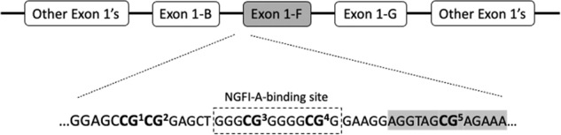
The figure shows the NR3C1 gene structure of exon 1 variants, as reported by McGowan et al. [13]. Below exon 1-F, we show part of the promoter and exon 1-F nucleotide sequences, with the five CpG sites analyzed in our study, denoted in bold (CG1, CG2, CG3, CG4, and CG5). Our CG1-CG5 sites correspond to CG35–CG39 sites analyzed in McGowan et al. [13]. The nucleotide sequence inside the broken box corresponds to a binding site of the nerve growth factor-induced protein A (NGFI-A) in the proximal promoter region of exon 1-F, as reported by McGowan et al. [13]. The gray-shaded area of the exon 1-F sequence indicates the beginning of the exon.