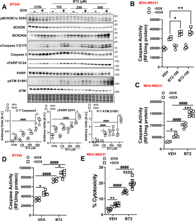
Fig. 3. BT2-mediated BCKDK inhibition induces cell death and potentiates DOX-mediated apoptosis in TNBCs.
A Immunoblot and densitometric analysis of total and phosphorylated BCKDE1α Ser 293, BCKDK, total and cleaved Caspase 3, total and cleaved PARP, total and phosphorylated ATM Ser 1981 in BT549 cells pre-treated with 125, 250, and 500 µM BT2 for 20 h followed by 2 µM DOX or DMSO treatment for 18 h. B Measurement of Caspase 3 activity in BT549 cells pre-treated with 100 µM and 150 µM BT2 for 20 h followed by 2 µM DOX or DMSO treatment for 18 h. Caspase 3 activity was measured in MDA-MB231 (C) and BT549 (D) cells and LDH release into the media was measured in MDA-MB231 cells (E) pre-treated with 500 µM BT2 for 20 h followed by 2 µM DOX or DMSO treatment for 18 h. Quantifications are from three independent experiments. A, *within groups (DMSO vs DOX), #between DOX groups (siCON vs siBDK#1 or vs siBDK#2); C–E, *within groups (DMSO vs DOX), #between groups (VEH vs BT2 and VEH+DOX vs BT2+DOX). Data presented as mean ± S.D. Statistical analysis was performed using a two-way ANOVA followed by a Tukey’s multiple comparison test; *p < 0.05, **p < 0.01, ****p < 0.0001 as indicated.

