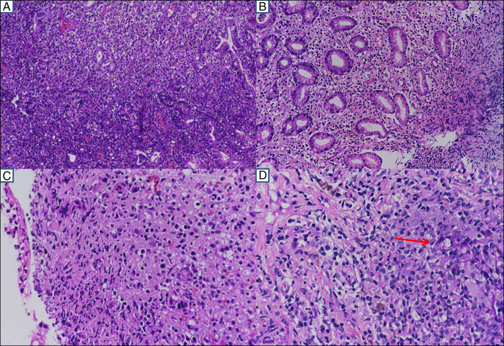
Figure 4.
(A) Diffuse infiltrate of atypical lymphoid cells displaying the gastric glands (20× magnification), (B) similar infiltrate with vacuolated cytoplasm (20× magnification), (C) ulcerated colonic mucosa with a similar type of infiltrate with slight nuclear irregularity, eosinophilic to vacuolated cytoplasm (40× magnification), and (D) the dermal infiltrates are seen infiltrating the rete pegs (red arrow) with similar nuclear and cytoplasmic features (40× magnification).

