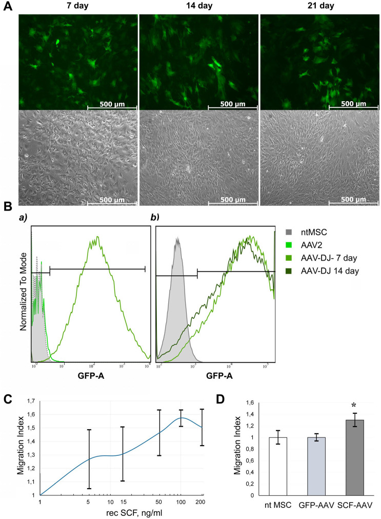
Fig. 3.
GFP expression efficiency and SCF functionality in rat MSCs after transduction with AAV-DJ. (A) AAV-DJ-infected rat MSCs exhibited robust GFP signal. Phase contrast (top) and green channel (bottom) representative images at 100x magnification. (B) Flow cytometry histograms showing GFP expression in rat MSCs transduced with AAV2 or AAV-DJ (a) and changes in GFP expression with AAV-DJ on the 7th and 14th days after infection (b). (C) Transwell migration assay of rat c-kit+ cardiac progenitors. Cells were allowed to migrate toward increasing concentrations of recombinant rat SCF in serum-free medium overnight. Five random fields of view were photographed for each sample, and number of migrated cells per FOV was counted. The cell migration index was calculated as the ratio of cells migrated toward SCF to cells migrated toward assay media. (D) Migration of rat c-kit+ cardiac progenitors toward conditioned media from the intact rat adipose tissue-derived MSC or transduced with GFP-AAV-DJ and SCF-AAV-DJ respectively in Transwell chamber. Data are expressed as the mean migration index±s.d. *P<0.05 versus ntMSC.

