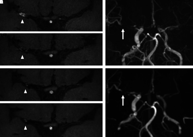
Fig. 2.
Hemorrhagic stroke case. A 50-year-old female presented with an intracranial hemorrhage at the left parietal lobe. Transverse time-of-flight MRA indicates right middle cerebral artery (MCA) stenosis and left internal carotid artery stenosis. The MRA and corresponding contrast high-resolution vessel wall MRI obtained at the initial evaluation (A), and 6 (B), 12 (C), and 24 (D) months later are provided. The intensity of vessel wall enhancement (VWE) at right MCA was changed (grade 2→grade 1→grade 1→grade 1; white arrowhead). The stenosis progression was observed at right MCA (E and F; white arrow).

