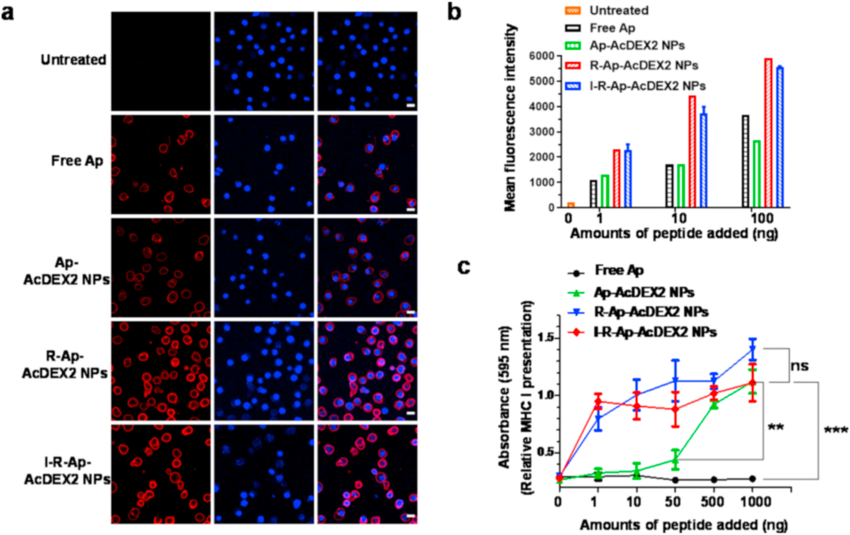
Fig. 4.

Detection of Ap presented by MHC-I of cancer cells. EL4 tumor cells (5 × 10⁵) were incubated with either free Ap, Ap-AcDEX2 NPs, I-R-Ap-AcDEX2 NPs, or R-Ap-AcDEX2 NPs for 6 h. (a) The resulting cells were spiked with PE-labeled anti-mouse H-2Kb/SIINFEKL antibody for 30 min, stained with DAPI, and imaged by confocal microscopy. Scale bars: 20 μm. (b) The cells were spiked with PE-labeled anti-mouse H-2Kb/SIINFEKL antibody for 30 min and analyzed by flow cytometry. (c) MHC-I antigen presentation (B3Z assay) by EL4 cells. In vitro CTL activation study of B3Z T cells co-cultured with EL4 cells following incubation with free Ap, Ap-AcDEX2 NPs, R-Ap-AcDEX2 NPs, or I-R-Ap-AcDEX2 NPs. The error bars show s.e.m. of three replicates. ns: no significant difference, **p < 0.01, ***p < 0.001. The p values are analyzed by two-way ANOVA (Bonferroni post-test) with GraphPad Prism.
