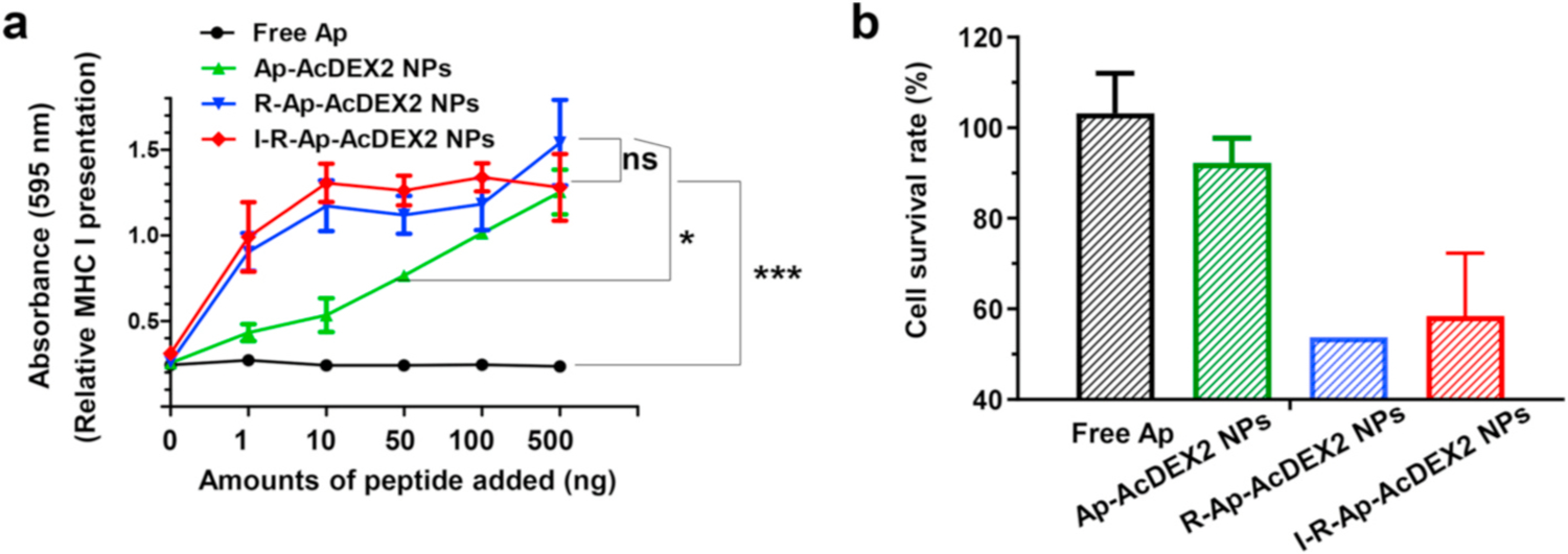
Fig. 5.

CTL activation studies. (a) MHC-I antigen presentation (B3Z assay) by BMDCs. In vitro CTL activation study of B3Z T cells co-cultured with BMDCs following incubation with free Ap, Ap-AcDEX2 NPs, R-Ap-AcDEX2 NPs, or I-R-Ap-AcDEX2 NPs. The error bars show s.e.m. of three replicates. ns: no significant difference, *p < 0.05, ***p < 0.001. The p values are analyzed by two-way ANOVA (Bonferroni post-test) with GraphPad Prism. (b) In vivo CTL activation study. Mice (1–2 per group) were subcutaneously immunized once with PBS, free Ap, Ap-AcDEX2 NPs, R-Ap-AcDEX2 NPs, or I-R-Ap-AcDEX2 NPs. 7-day later, CFSEhi labeled Ap pulsed target cells were injected into the immunized mice together with CFSElo labeled control cells. 24 h after cell injection, mice were euthanized, and their splenocytes were prepared for FACS analysis.
