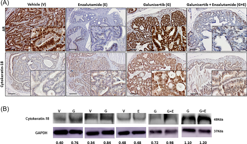
FIGURE 7.
TGF-β blockade induces prostate tumor cell differentiation independent of AR. Panel A shows representative characteristic images of cytokeratin-18 immunoreactivity and AR expression and nuclear localization in prostate tumor sections. Upper panel, reveals AR immunoreactivity and lower panel cytokeratin-18 immunoreactivity in serial sections of prostate tumors after various treatments, vehicle control (V), enzalutamide monotherapy (E), galunisertib monotherapy (G), and the combination treatment of galunisertib and enzalutamide (G+E) (dosing as described in section 2 for 2 weeks). Galunisertib monotherapy led to a marked increase in cytokeratin-18 immunoreactivity indicating tumor re-differentiation, compared to the enzalutamide alone and vehicle control (Magnification ×200, insert ×1000). Panel B indicates the cytokeratin-18 expression profile in response to various treatments as detected by Western blot analysis. The results shown are representative of three independent experiments for each of the three “mini”-trials. Protein expression was determined by densitometric analysis of band intensity (numerical values shown at the bottom of blot). Galunisertib alone (G) resulted in approximately twofold increase in cytokeratin-18 expression, compared to vehicle controls (V), while there was no apparent difference with enzalutamide alone (E). The combination of galunisertib and enzalutamide (G+E) had no major effect on cytokeratin-18 levels.

