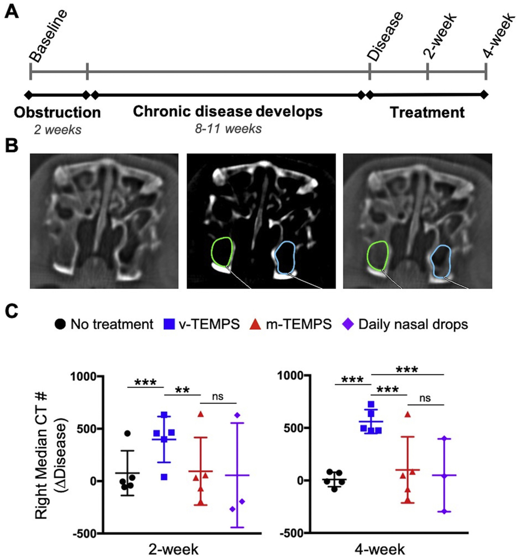
Fig. 3.

MicroCT imaging showed reduced opacification of the right maxillary sinus following application of m-TEMPS compared to v-TEMPS. (A) Disease was induced by creating a reversible obstruction of the left ostiomeatal complex for 2 weeks. Over the subsequent 11 weeks (disease induction) or 8 weeks (disease re-induction) chronic disease phenotype developed [16]. Subjects were divided into 4 groups for bilateral treatment and monitored for 4 weeks. (B) Representative coronal microCT images of the maxillary sinuses in soft tissue and bone (center) windows. Using the bone window, opacification was measured in a defined region of interest and the median CT # of that region was recorded for 3 consecutive sections. (C) Change in CT # between the disease state and treatment timepoints on the right side. Error bars represent the mean ± S.D. for each group: no treatment (n = 5), v-TEMPS (n = 5), m-TEMPS (n = 5), and daily nasal drops (n = 3). Statistical significance was determined by one-way ANOVA with post hoc Wilcoxon Method testing, ***p < 0.001, **p < 0.020, ns = not significant.
