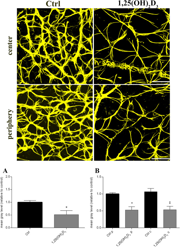
Fig. 7.
The 1,25(OH)2D3 treatment reduced reactive astrocytes in DBA/2J mice. Upper panel shows representative images GFAP+ astrocytes assessed in flat-mount retina, of control (vehicle-treated) and 1,25(OH)2D3-treated group. Scale bar, 50 μm. The density of activated astrocytes was quantified, measuring the mean grey levels of GFAP staining density. Quantification was carried out on whole retina (A), on both retinal centre and periphery (B). Data are plotted as mean ± SD (n = 6 flat-mount retinas for each experimental group, n = 48 images for each experimental group): A *p < 0.0, p = 0.0142 vs. Ctrl, t-test two-tailed. B *p = 0.0029 vs. Ctrl periphery (Ctrl p) ‡p = 0.0029 vs. Ctrl centre (Ctrl c), one-way ANOVA with Tukey post hoc test for multiple comparisons

