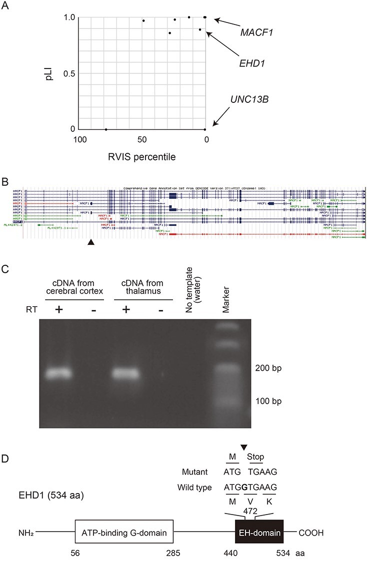
Figure 1 .

De novo LOF mutations reported in patients with bipolar disorder. (A) Plot of RVIS percentile and pLI of the genes in which de novo LOF mutations were previously found (13). Arrows show the genes coding calcium binding proteins (MACF1, EHD1 and UNC13B). (B) Structures of MACF1 isoforms. An arrowhead indicates the location of the de novo mutation on a minor exon. Data are retrieved from the UCSC database. (C) RT-PCR of the minor isoform of MACF1. The minor exon of MACF1 in which the de novo mutation was found was amplified using cDNA derived from human cerebral cortex and thalamus. RT, Reverse-transcription. (D) Schematic diagram of the domain structure of EHD1 and the location of the de novo mutation of EHD1. The mutation, 1-bp deletion, was found in the last exon, and a truncated protein lacking most of the EH-domain is predicted to be expressed. aa, amino acids.
