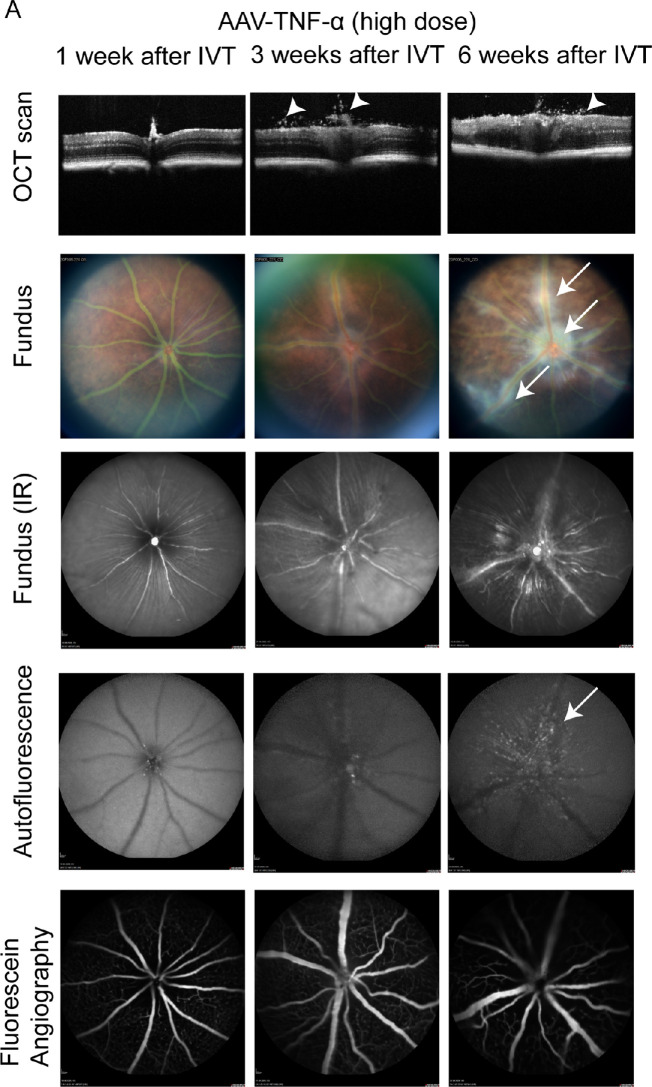
Figure 4.
Inflammation was observed in AAV-TNF-α injected eyes. (A) OCT scans revealed cellular infiltrates in the vitreous (white arrowheads) 3 to 6 weeks after injection of AAV-TNF-α (upper row). White cellular infiltrates were observed around the vasculature and the optic nerve on fundus pictures (second and third row, arrows) in AAV-TNF-α injected eyes. Few hyperfluorescent spots were found around the optic nerve and vasculature in AAV-TNF-α injected eyes by BAF. FFA was largely normal in AAV-TNF-α eyes. Different animals were examined at 1, 3, and 6 weeks after IVT. 1 × 10⁹ VG/eye AAV-TNF-α was injected intravitreally.

