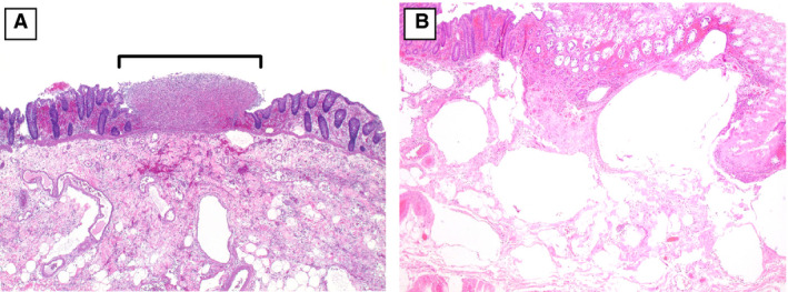
Figure 2.

A, A segment of colon with a discrete, punctate area of necrosis (bracket) adjacent to viable mucosa [haematoxylin and eosin (H&E)]. B, Markedly oedematous submucosa with numerous large empty spaces, consistent with pneumatosis intestinalis (H&E).
