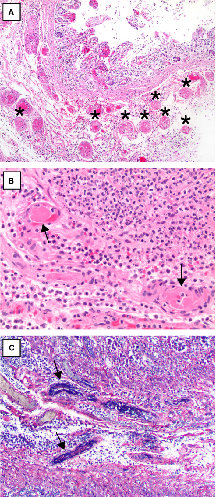
Figure 3.

A, A segment of small intestine with numerous submucosal small vessel fibrin thrombi (asterisks) beneath an area of necrotic mucosa [haematoxylin and eosin (H&E)]. B, A higher‐power view of two well‐formed submucosal fibrin thrombi (arrows) (H&E). C, Phosphotungstic acid haematoxylin staining highlights fibrin thrombi in small vessels (arrows).[Colour figure can be viewed at wileyonlinelibrary.com]
