
FIGURE 2.
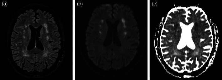
Axial fluid‐attenuated inversion recovery (FLAIR) magnetic resonance image (a), axial diffusion‐weighted image (b) and apparent diffusion coefficient map (c) of the brain. Bilateral multiple, small, symmetric FLAIR hyperintensities (a) that are linearly oriented parallel to the lateral ventricles in the corona radiata showing diffusion restriction (b–c), indicating deep watershed infarcts