
FIGURE 4.
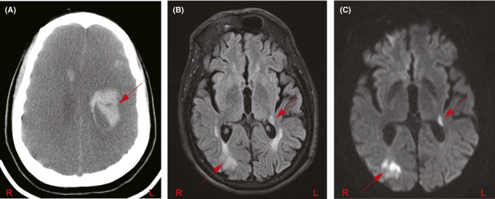
Neurological complications in COVID‐19 patients. (A) Non‐contrast CT head shows diffuse loss of gray‐white differentiation and sulcal effacement, consistent with global hypoxic ischemic encephalopathy. Additionally, there are multifocal intracranial hemorrhages, including a large hematoma in the left fronto‐parietal deep white matter (red arrow); (B) T2‐FLAIR and (C) DWI MRI brain sequences in a patient show right parieto‐occipital and left posterior thalamocapsular acute infarcts (arrows)