Figure 3.

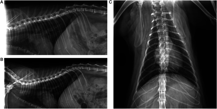
2D thorax radiograph result of the patient to examine the potential pathological results of the SARS‐CoV‐2 infections. (A) Right lateral thorax graphy, (B) left lateral thorax graphy, (C) ventral‐dorsal thorax graphy.

2D thorax radiograph result of the patient to examine the potential pathological results of the SARS‐CoV‐2 infections. (A) Right lateral thorax graphy, (B) left lateral thorax graphy, (C) ventral‐dorsal thorax graphy.