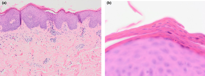
Figure 2.

(a) Prominent alternating orthokeratosis and parakeratosis in horizontal and vertical directions. The epidermis showed mild irregular acanthosis with broader rete ridges than expected in a psoriasiform reaction. There was mild and focal spongiosis with slight lymphocytic exocytosis. There was also mild perivascular and perifollicular lymphocytic inflammation within the papillary dermis with neutrophils and occasional eosinophils seen focally. (b) Prominent alternating orthokeratosis and parakeratosis in the distinct ‘checkerboard’ pattern. Haematoxylin and eosin, original magnification (a) × 20; (b) × 600.
