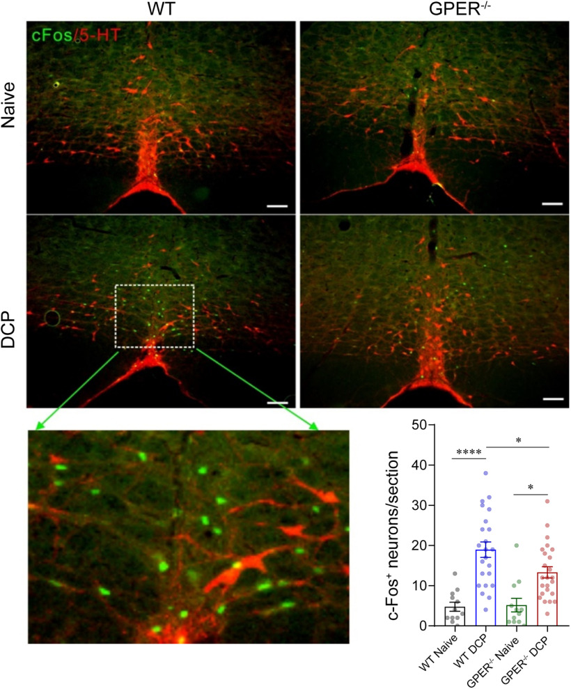
Figure 7.
GPER deficiency results in fewer activation of RVM neurons in contact dermatitis female rat models. Representative IHC images and graph showing c-fos+ neurons and serotonergic neurons in the RVM of naive and DCP-treated WT and GPER−/− rats. Scale bar, 100 µm. The scatter plots show number of c-fos+ neurons in 12 slides from three rats for naive groups and 23–24 slides from five rats for DCP-treated groups. Differences were compared using one-way ANOVA and Tukey's post hoc tests. F = 15.10; p < 0.0001 for WT naive versus WT DCP, p = 0.0445 for WT DCP versus GPER−/− DCP, p = 0.0106 for GPER−/− naive versus GPER−/− DCP.

