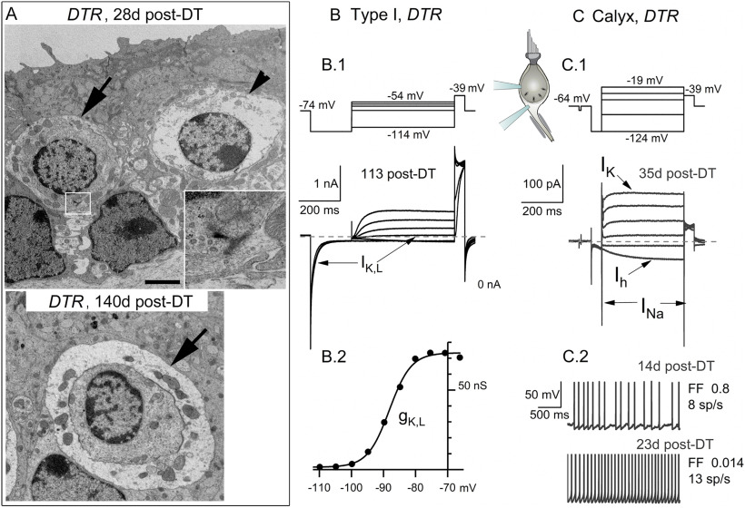
Figure 11.
A small number of type I hair cells and calyces in DTR utricles survived DT treatment and appeared normal. A, TEM of healthy-appearing type I hair cells and calyces (arrows) at early (top) and later (bottom) times post-DT treatment. A presynaptic ribbon (box, top) is magnified in the inset. Top, Arrowhead, Type I hair cell undergoing cytolysis. Scale bar: top, 2.5 µm; (in top) bottom, 2.2 µm. B, C, Whole-cell voltage-gated currents from surviving type I hair cell (B.1) and calyx terminal (C.1) in DTR utricles. Voltage protocols (top) evoked currents (shown below voltages) with normal time course and voltage dependence. B.2, G(V) relationship for cell in B.1, fit with Equation 1 (Boltzmann function) with V1/2 = –78 mV, S = 6 mV, Gmax = 47 nS, which are consistent with normal values for gK,L. C.2, Spontaneous spiking in two calyceal terminals with different rates [spikes per second (sp/s)] and regularity of spike intervals [measured by Fano Factor (FF)].

