Figure 3.
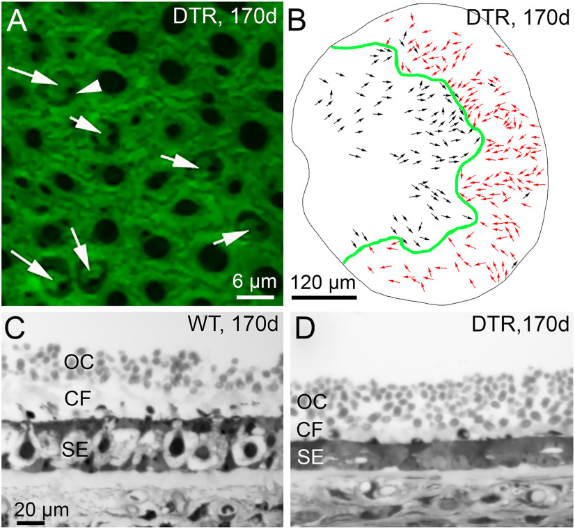
The map of hair bundle orientations and the otoconial layer appeared normal in regenerated DTR utricles. A, Hair bundle orientation was visualized by phalloidin labeling of filamentous actin at the apical surface of the utricular macula. A dense network of F-actin in the junctional complexes of supporting cells is punctuated by holes that, in six cases (white arrows), reveal the actin-containing cuticular plates of regenerated hair cells. Arrowhead points to the black hole (lack of stain) at the site of the microtubular kinocilium to one side of a stereociliary bundle. White arrows show the orientations of each hair bundle (see text). B, Map of regenerated bundle orientations for one DTR utricle: green line, LPR; black arrows, orientations pointing from medial to lateral (left to right); red arrows, orientations pointing lateral to medial. Most orientations were expected for their location relative to the LPR; several black arrows lateral to (right of) the LPR show exceptions. C, D, Transverse sections through WT (C) and DTR (D) utricles at 170 d post-dT show otoconia crystals (OCs) and column filament (CF) material above the sensory epithelium (SE). Scale bar: C (for C, D), 20 μm.

