. 2021 May 28;81(16):4305–4318. doi: 10.1158/0008-5472.CAN-20-3929

Figure 3.

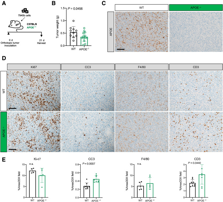
Figure 3. Loss of APOE results in reduced tumor burden and fibrosis. A, Experimental scheme for orthotopic transplantation of 7940b, KPC tumor cells. B, Final tumor weight (g) in WT (n = 10) and ApoE–/− (n = 13) mice. Statistical significance was determined using two-tailed t test, with a P < 0.05 considered statistically significant. C, Representative IHC for APOE in WT and ApoE–/− mice. Scale bar, 100 μm. D, Representative IHC staining for Ki-67, cleaved caspase-3 (CC3), F4/80, and CD3 in WT and ApoE–/− mice. Scale bars, 100 μm. E, Quantitation of IHC stain as a percentage area per 20× field in WT (n = 4–8) and ApoE–/− mice (n = 5–8). Statistical significance was determined by two-tailed t tests. n.s., not significant.

Loss of APOE results in reduced tumor burden and fibrosis. A, Experimental scheme for orthotopic transplantation of 7940b, KPC tumor cells. B, Final tumor weight (g) in WT (n = 10) and ApoE–/− (n = 13) mice. Statistical significance was determined using two-tailed t test, with a P < 0.05 considered statistically significant. C, Representative IHC for APOE in WT and ApoE–/− mice. Scale bar, 100 μm. D, Representative IHC staining for Ki-67, cleaved caspase-3 (CC3), F4/80, and CD3 in WT and ApoE–/− mice. Scale bars, 100 μm. E, Quantitation of IHC stain as a percentage area per 20× field in WT (n = 4–8) and ApoE–/− mice (n = 5–8). Statistical significance was determined by two-tailed t tests. n.s., not significant.