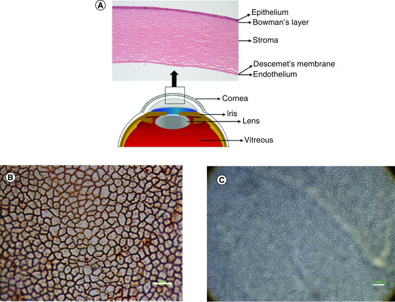
Figure 1. . Structural anatomy of the human cornea and corneal endothelial cells.
(A) The cornea consists of five layers; epithelium, Bowman's layer, stroma, Descemet's membrane and endothelium. (B) Corneal endothelial cells presented hexagonal morphology after staining with alizarin red. (C) Corneal endothelial cells observed under light microscope.
Scale bar = 100 μm.

