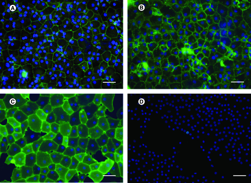
Figure 2. . Immunocytochemistry using different biomarkers.
Human corneal endothelial cells were stained in green for (A) ZO-1, (B) 1A3, (C) 2A12 and (D) Ki-67. Nuclei were counterstained in blue. Hexagonal shape of corneal endothelial cells was clearly observed after stained with cell surface marker antibodies. Cell proliferation marker, Ki-67, was used to evaluate number of cells which have proliferative capacity.
Scale bar = 100 μm.

