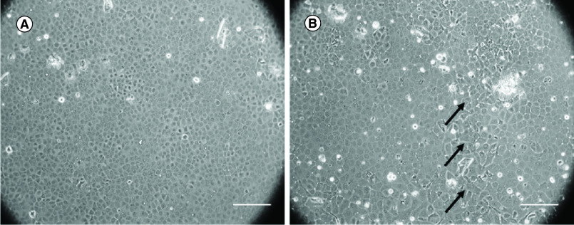
Figure 3. . Human corneal endothelial cells from young and older donors.
(A) Human corneal endothelial cells from the young donor showed homogeneous hexagonal morphology. (B) Human corneal endothelial cells from older donor showed polymegathism and pleomorphism. The polymorphic cell is marked with black arrows.
Scale bar = 250 μm.

