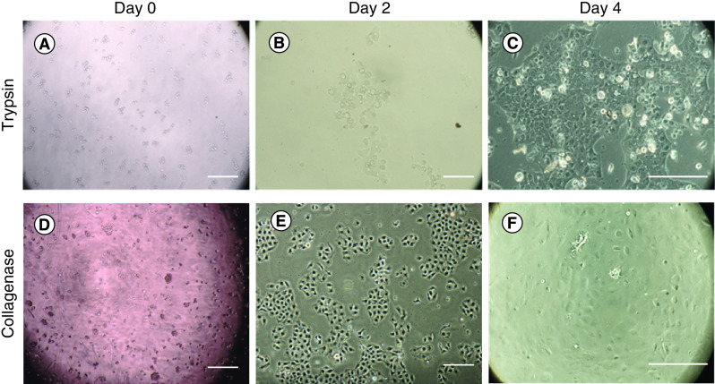
Figure 4. . Human corneal endothelial cell isolation using trypsin and collagenase.
At day 0, (A) the cells digested with trypsin presented smaller cell number compared with (D) collagenase. At day 2, (E) the cells digested with collagenase showed better proliferation than the cells digested with (B) trypsin. At day 4, (F) nearly 50–70% confluence was found in the collagenase group. Meanwhile, (C) around 30–40% cell confluence was found in trypsin group. Scale bar = 250 μm.

