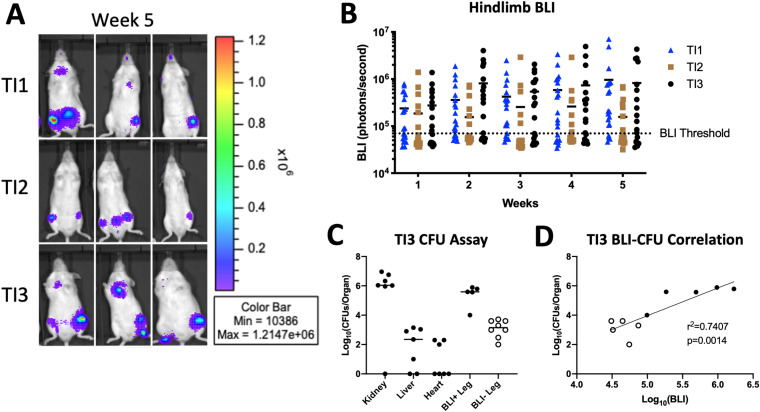
FIG 3.
Characterization of hematogenous osteomyelitis using clinical isolates of S. aureus. (A) In vivo bioluminescence imaging (BLI) of three representative mice for each isolate at 5 weeks postinjection. (B) Quantification of the BLI signal measured in the hindlimb of each injected mouse depicted over the 5 weeks postinjection (n = 20 per isolate) (the BLI threshold is 70,000 photons/s). (C) Enumerated bacterial burdens from the indicated organs, as measured by a CFU assay at 3 weeks postinjection. BLI+ and BLI− legs are the counts from femurs from legs with BLI signals higher and lower than 70,000 photons/s, respectively (n = 7 mice from a separate cohort from those in panels A and B injected with TI3) (each dot represents one leg). (D) Correlation of the bacterial burden (CFU) from panel C with the BLI signal of the corresponding leg. Open and closed dots correspond to the BLI+/− designations in panel C.

