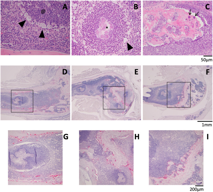
FIG 6.
Detailed histological characteristics of HOM. (A and B) Abscesses without (@) (A) and with (*) (B) visible bacterial colonies were surrounded by enlarged macrophages (black arrowheads). (C) Magnified image of the sequestrum from Fig. 5F showing bacteria (black arrows). (D to I) TRAP staining (red) for osteoclast activity. Panels G to I depict higher magnifications of the black boxes indicated in panels D to F, respectively. Scale bars are presented for each row of images.

