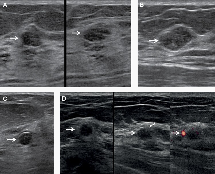
Figure 1.
Images of a 57-year-old woman with multiple bilateral masses on screening US, including cysts, presumed fibroadenomas, and invasive ductal carcinoma with ductal carcinoma in situ (IDC-DCIS). Oval, circumscribed hypoechoic masses (arrows) are seen in the left breast at 2 o’clock (A) (left, radial; right, antiradial) and the right breast at 2 o’clock (B) with no posterior features, typical of BI-RADS 3, probably benign masses on screening US. These can be followed at one year, and were both stable at subsequent 12- and 24-month screening US examinations. A benign rim-calcifying cyst (C, arrow) was seen in the right breast at 12 o’clock. Multiple simple cysts were also present (not shown). A hypoechoic oval mass (arrows) with a subtle echogenic rim on the radial image and focally indistinct margin (curved arrow) was seen in the left breast at 10 o’clock (D) (left, radial; center, antiradial; right, power Doppler). This is a BI-RADS 4A, low suspicion mass, which underwent targeted US then core-needle biopsy and excision showing a 1.2 cm grade 2 IDC-DCIS, estrogen receptor positive, progesterone receptor weakly positive, human epidermal growth factor 2 negative, with negative sentinel node biopsy.

