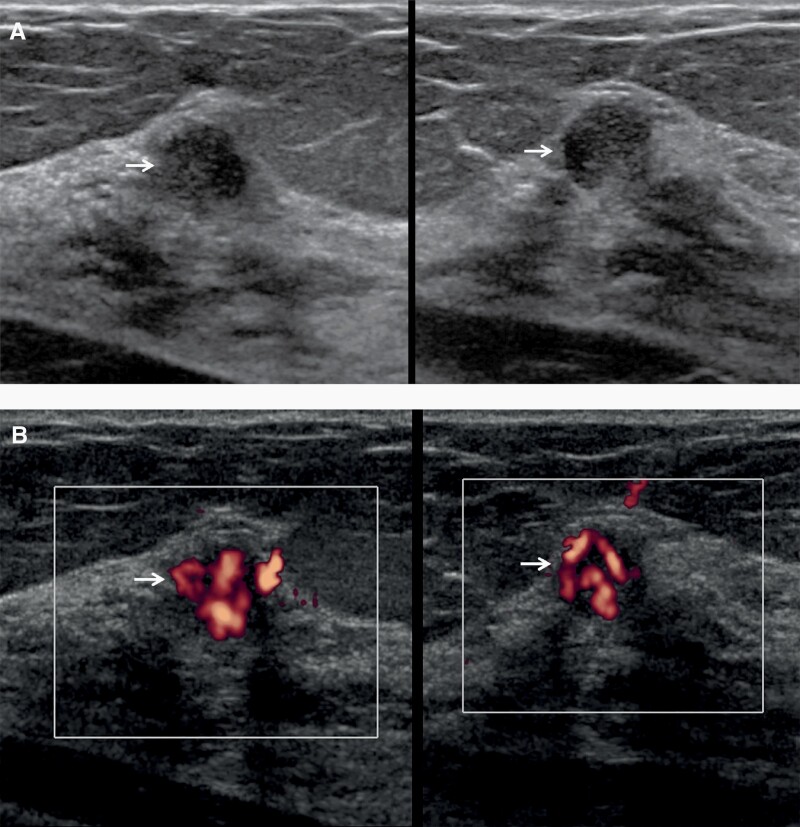
Figure 4.
Images of a 68-year-old woman with technologist-performed screening US-detected cancer. A: Radial (left) and antiradial (right) US images show a round hypoechoic mass (arrows) with partially circumscribed and partially indistinct margins. Strong internal vascularity was evident in the mass on power Doppler images (B, arrows), a suspicious finding. This was assessed as BI-RADS 3 (with recommendation for 6-month follow-up) by one radiologist and recommended for immediate additional imaging by the second radiologist as part of a research protocol. At targeted US, it was assessed as BI-RADS 4A, low suspicion. US-guided core-needle biopsy showed atypical ductal hyperplasia and papilloma, upgraded to nuclear grade 2 ductal carcinoma in situ at excision.

